Atrial Fibrillation? Better ask your doctor about anticoagulation.

4ipw7

Atrial fibrillation (AF) is a major risk factor for TIA and Stroke.

It is estimated that AF accounts for >20% of all strokes.

This risk can be lowered by as much as 60% by taking an anticoagulant such as warfarin (Coumadin).

Current guidelines recommend anticoagulation for all patients with AF, even paroxysmal (intermittent) AF:

1. If they have had a prior TIA or stroke, or

2. If they have two or more of the following risk factors: (1) age > 75 years, (2) history of hypertension, (3) diabetes mellitus, and (4) moderately or severely impaired left ventricular systolic function and/or heart failure.

If you have AF, click here to calculate your CHADS2 score and stroke risk, and click here to calculate your risk of bleeding from anticoagulation.

Your risk of stroke is much higher than your bleeding risk, right?

Many AF patients don’t get anticoagulants because they are considered a fall risk. If you had AF and need anticoagulation, you would have to fall more than 300 times a year for the harm from the falls to outweigh the benefits of anticoagulation.

Despite these guidelines, recent studies have shown that:

1. Less than half of AF patients with a high stroke risk receive anticoagulants.

2. Even less patients with paroxysmal (intermittent) AF than those with permanent AF receive anticoagulants (31 vs 49%), even though the stroke risk is the same in both groups.

3. Too few patients with new onset AF are started on anticoagulants (only 52%).

We need to do better.


Click here to download a booklet about AF and stroke.

Click here to download a worksheet that you and your doctor can use to decide if anticoagulation is right for you.

Teleneurology – Get specialist care faster!

telemed1

We have already blogged about the importance of getting thromolytic therapy as quickly as possible for acute stroke.

ambulance

We have urged you to call 911, and get to the hospital by Ambulance.

Certified stroke centers, like Monmouth Medical Center, have established protocols to get brain scans and blood work done as quickly as possible.

But what about if the Neurologist can’t get to the hospital quickly enough?

jwj Obama 00510

Suppose it’s late in the evening on July 4th, and the Neurologist gets stuck in traffic driving to the hospital.

time is brain

You’re sitting in the emergency room with a stroke losing precious minutes.

Teleneurology allows the Neurologist to use a secure video conferencing system to examine and counsel an emergency room patient from wherever he or she may be:

Find out more about the benefits of teleneurology.

Monmouth Medical Center is currently investing in telemedicine and teleneurology to improve patient care.

Watch this space for more information!

Most stroke patients are not getting treated with t-PA!

alteplase-500x500

We have already blogged about acute stroke, thrombolytic therapy with t-PA, and the importance of getting to the hospital right away for early treatment.

xx

However, even now that t-PA can be administered to most stroke patients within 4.5hrs since the onset of their symptoms, only around 5% of acute stroke patients receive the clot busting drug in the USA, more (7-8%) at certified stroke centers.


The #1 reason for not receiving t-PA is missing the time window for safe administration.

The drug must be administered within 4.5hrs (or 3hrs for some patients) since the onset of stroke symptoms in order for the benefits of the drug to exceed the risk.

Why don’t stroke patients get to the hospital in time?

Uncertain time of stroke onset.

In many cases, stroke happens during sleep, patients go to bed normal and wake up many hours later with a stroke.

We don’t know exactly what time the stroke occurred, and we have to go by the time when they were last known to be normal, which often put them outside that 3-4.5hr window.

Lack of knowledge.

However, there are still many patients who could get to the hospital in time to receive t-PA but don’t.

This is a failure of public education.

Studies have shown that as many as 1/3 of people surveyed cannot name a single symptom of stroke, and that 1/10 of people surveyed are not aware there is a time sensitive treatment available.

Only 2/3 stoke patients choose to call 911 and come to the hospital by ambulance – those that do are 50% more likely to get t-PA.

Stroke

time is brain

This is the reason for the ad campaigns like the AHA’s “act FAST” and “Time is brain”.


Even if they do get to the ED in time, most stroke patients still don’t get t-PA

Only 1/3 of acute stroke patients who do get to the hospital in time get t-PA!

Many have a definite contraindication, like taking blood thinners or having had recent major surgery, that does make the treatment too risky.  These are a subset of stroke patients who should be considered for interventional procedures to directly retrieve blood clot from the cerebral arteries at comprehensive stroke centers.

However, as many as a 1/3 of acute stroke patients seen within that 3-4.5hr time window do not receive t-PA because their neurologic deficit is considered to be “too mild or rapidly reversing”.  Obviously, this is a statement that is so hard to define, and yet still considered by many physicians to be contraindication to the use of the drug.

Many of us would argue that to the patient, there is no such thing as  a “mild” stroke.

Image 1 in Holland-TREC

Some affected patients just get a quick screening examination lying on a gurney, and are quickly dismissed as having had “mild stroke” even though they can’t stand and walk because this was never tested.

Furthermore, there is increasing evidence that patients who truly have minimal stroke symptoms can still benefit from t-PA administration.

!cid_8D464BF3-277A-4528-BC41-3735380B61DB

Our own research from our certified stroke center at Monmouth Medical Center has corroborated these concerns:

We reviewed the charts from all acute stroke patients seen at Monmouth Medical Center from 2008-2012:

8% got t-PA.

75% were excluded because came outside the 3-4.5hr time frame for administration of t-PA.

9% were seen in time for t-PA, but either refused treatment or couldn’t get it because if a definite contra-indication.

8% did not get t-PA because of a “mild or rapidly improving deficit” – of those, 13% needed rehab placement, so there deficit wasn’t so mild after all!

xx

Obviously, our goal is to give more t-PA to stroke patients who can benefit from the clot busting treatment.

We are working hard on public education events to get stroke patients to come to ED sooner, and we are also going to be treating more patients with “mild” deficits as long as they meet eligibility criteria.

xx

What can you do?

Know the warning signs of stroke.

If you, or anyone you know, shows any of these signs call 911 and get them to the hospital right away.

Be a participant in your own or your loved one’s medical decision making – if there seems to be any residual stroke symptoms (however mild) ask about t-PA treatment for stroke.

Find out more about Monmouth Medical Center’s certified stroke center here.

Correct clinical diagnosis of dizziness in the ER could save $$ billions

vertigo_dizziness-150x150
We have already blogged about the danger of escalating health costs in the USA.

The cost of emergency room visits for severe dizziness has grown exponentially in recent years, topping $3.9 billion in 2011 and projected to reach $4.4 billion by 2015.

Investigators from Johns Hopkins estimate that half a billion a year could be saved immediately if emergency room physicians stopped the routine and excessive use of head CT scans to search for stroke in dizzy patients, and instead used simple bedside physical exams to identify the small group of patients that truly needs imaging.

Click here to find out more about the Johns Hopkins study.

Click here to find out more about the hidden dangers of unnecessary CT scans.

Once they understand how inner ear disease can cause vertigo and nystagmus, any physician can use our simple clinical scheme to distinguish inner ear problems from more serious and rarer central causes like stroke in dizzy patients.  Click here to find out how.

A ripping yarn – a tale of cervical artery dissection

Case Summary:
This 46-year-old woman was healthy except for a history of occasional migraine headaches and cigarette smoking. On the day of admission she had fallen down a short flight of steps carrying a heavy box. About 2 hrs later she complained of some neck pain.  Then later that evening developed abrupt onset left sided weakness. She arrived at the emergency room within 1.5 hrs of the onset of weakness. On examination, she was alert, but she had a right gaze deviation (she wouldn’t look to the left side) and the left side was completely paralyzed. She had a normal brain CT scan.

The stroke team was notified, and she was given intravenous thrombolytic (“clot busting”) drug therapy within 1/2 hr of her arrival at the hospital and 2 hrs since the onset of her symptoms.

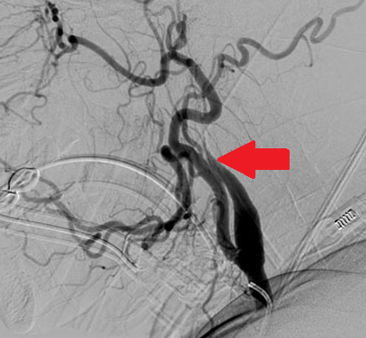
Carotid ultrasound subsequently showed no flow in the right internal carotid artery, and carotid arteriography subsequently showed near occlusion of the artery from an arterial dissection (see image below, red arrow):

dissection2

xx

What is cervical artery dissection?

Cervical artery dissection is caused by bleeding inside the wall one of the major arteries in the neck.

This process is thought to be triggered by local injury to the inside layer of the vessel wall.

Cervical artery dissections occur from blunt trauma:

dissection4

Cervical artery dissection can also occur after minor trauma, particularly in someone with a genetic predisposition:

dissection5

xx

What diseases predispose to arterial dissection?

There are some specific syndromes such as Marfan syndrome, Pseudoxanthoma elasticum and Ehlers- Danlos syndrome type IV that are associated with a weakness in the arterial wall making an arterial dissection more likely:

dissection6

dissection7

dissection8

In other cases, the specific cause of arterial weakness is unknown, but there is ongoing research to try to identify genetic links.

xx

What happens after a cervical artery dissection?

Symptoms can be caused from the damaged arterial wall itself (local symptoms) and some affected patients will later develop strokes.

Local symptoms include neck pain, unusual headache and/or Horner’s syndrome.

dissection9

L Horner’s syndrome (small pupil and drooping eyelid) caused by damage to the sympathetic nerve fibers in the arterial wall from carotid dissection. Click here to find out more about Horner’s syndrome and other causes of unequal pupils.

xx

What about stroke?

Stroke symptoms only occur in 25-30% dissections and can occur several days after the neck trauma and/or onset of local symptoms.

The arterial dissection narrows the space inside the blood vessel (the lumen), so less blood flow gets to the brain:

dissection1

A carotid artery dissection with blood clot inside the arterial wall (left) leading to narrowing of the vessel lumen and less blood flow (right).

Cervical arterial dissections can also cause stroke when pieces of blood clot break off and move with the blood flow only to block small arteries further inside the brain (cerebral thromboembolism), or if the dissection tracks across (and blocks off) an arterial branch (see below):

dissection3

xx

How is arterial dissection diagnosed?

Magnetic resonance imaging is probably the easiest way to make the diagnosis:

dissection10

MR angiogram (left) showing tapered occlusion of the left internal carotid (white arrow) from dissection. Fat suppressed T1 weighted MR axial image through the dissected cervical artery (right) showing bright blood within it’s wall (black arrow) from dissection.

xx

How is it treated?

In most cases the arterial dissection ultimately heals on its own without any surgical intervention.  There has been some controversy surrounding the use of anticoagulant vs anti-platelet drugs for stroke prevention after cervical artery dissection, but most current data favors the use of the anti-platelet drug aspirin:

dissection11

Of course, for patients presenting with symptoms of acute stroke, throbolytic therapy is also an option, and can improve outcome without increased risk in stroke from dissection:

dissection12

Click here to find out more about cervical artery and dissection and stroke.

Click here to find out what to do if you think your having a stroke.

Click here to find out more the certified stroke center at Monmouth Medical Center.

Foreign Accent Syndrome – Their “Problem” or Yours?

FAS3

Foreign accent syndrome (FAS) is a rare condition which causes affected patients to suddenly speak their native language in a foreign accent.

Cases of FAS were reported as early as 1900.  However, one of the best known historical cases is “Astrid L”, a Norwegian woman who suffered a traumatic brain injury from shrapnel during a WW2 air raid in 1941.  She survived, but found herself mispronouncing vowels in such a way that she seemed to have a German accent, leading to social isolation and stigmatization for the remainder of the war.

Since then, there have been about another 60 FAS cases reported in the literature and media, mostly in patients who have suffered acute neurologic events such as strokes, multiple sclerosis and head injury.

Unlike most neurologic syndromes, FAS has not been localized to a lesion in a particular brain area.

The only thing that can be said is that most affected patients have lesions affecting the dominant hemisphere in or around known language areas.

FLS

Brain imaging studies from a FAS patient: The MRI (left) shows enlargement of the Sylvian fissure from atrophy of the left temporal lobe. The PET scan (right) shows focal hypometabolism in the left temporal lobe.

Many affected patients were initially mute, then developed FAS as they recovered from a non-fluent aphasia:

There are also some cases of FAS that have developed after minor neurologic events, or even without any clearly identifiable neurological cause at all.  Some of these patients have had normal brain imaging, suggesting that the problem can be functional or psychogenic.

This is all further complicated by the fact that different listeners can perceive different accents in a single speaker.

The video clip is a patent with FAS syndrome after brain injury from hemiplegic migraine.  She is said to have a Chinese accent.  Does it sound Chinese or just slurred to you?

The table below is from a FAS case report, where the affected patient’s “foreign accent” was obviously described very differently by observers.

FAS2

This suggests that FAS may not be a true syndrome after all, but simply a listener-bound epiphenomenon.

What does this mean?

Well, we have already explained that most FAS patients have some kind of speech or language problem that changed how they speak.  That explains the association with lesions in the dominant hemisphere.   However the “foreign accent” may actually just something perceived by the listener – the variability of perceived accents is explained by the fact that listeners have different experiences with languages other than their own.

In other words FAS may not be a true syndrome, but simply an epiphenomenon that exists only in the ears of the beholder.

What’s best for stroke prevention, aspirin, clopidogrel or both?

We have already blogged about the benefits of anti-platelet agents in stroke prevention.

We haven’t talked about how they work, or which one(s) are best.

Platelets are an important component of blood clotting (or hemostasis) – the normal process that stops us bleeding and facilitates healing after a cut or other injury:

This same platelet led coagulation cascade can lead to blood clot formation inside intact but diseased arteries laden with atheroma – the result of years of hypertension, high cholesterol and smoking:

These small blood clots then break off and travel down the artery causing (in the case of a cerebral blood vessel) a TIA or stroke.

There are three currently available  anti-platelet medications  – aspirin, modified release dipyridamole with aspirin (Aggrenox) and clopidogrel (Plavix) which inhibit platelet activation and aggregation.

asa

Comparative studies have shown that all three drugs reduce the risk of ischemic stroke in high risk patients who have had a previous stroke or TIA.  Aspirin is the cheapest of the three.  Aggrenox is more effective than aspirin alone, but can cause troubling headache in some patients.  Clopidogrel can be used in patients who are allergic or immune to aspirin.

Because these drugs work in different ways, investigators have asked whether combining them might be even more beneficial.

The initial studies said No.  The MATCH (2004), CHARISMA (2006) and SPS3 (2012) trials all showed that long term use of the combination of aspirin and clopidogrel failed to reduce the risk of major vascular events and also led to significant increased life-threatening bleeding complications (mainly intracranial and gastrointestinal) compared with either drug alone.

match

However, new data from the CHANCE study presented at this year’s international stroke meeting shows that a short course of combined treatment might be helpful:

The study enrolled 5170 patients who had suffered a TIA or minor stroke within the previous 24hrs, randomly assigned to one of two treatment groups:  The first group received aspirin (75-300 mg one-day loading dose followed by 75 mg/day).  The second group received the same aspirin regimen plus clopidogrel (loading dose of 300 mg followed by 75 mg/day) for 21 days, then just the clopidogrel alone after that.

The study showed that the 90d stroke incidence was lower in those who received both aspirin and clopidogrel.

The risk of hemorrhagic stroke and other severe bleeding was the same in the two groups.

In other words, short term combination anti-platelet therapy might be more effective in preventing stroke in this high risk TIA and minor stroke group, and this is something we will be offering patients seen in Monmouth’s innovative TIA Rapid Evaluation Center.

Magnetic Brain Stimulation Might Help Stroke Rehab?

Repetitive transcranial magnetic stimulation (rTMS) of the brain has been used to treat a variety of neurologic and psychiatric disorders including depression and dytonia.

A new study published this week suggests that it might also help speed recovery of speech and language in stroke survivors.  The  study included 24 stroke survivors with aphasia. Thirteen of them received transcranial magnetic stimulation (TMS) for 20 minutes every day for 10 days followed by speech therapy.  The remaining 11 received a “sham” brain stimulation.

Patients in the TMS group showed three times greater improvement than those in the sham stimulation group.

Find out more here.

Dr Holland and Monmouth Neuroscience Institute’s TIA Rapid Evaluation Center Honored at the 2013 Heart Ball

The American Heart Association and American Stroke Association gathered to recognize Dr. Neil Holland and Dr. June Duck as this year’s medical honorees at the annual Heart Ball on June 21 at the Ocean Place Resort and Spa in Long Branch.

IMAG0529

Drs Holland and Duck with their awards.

Dr. Holland was recognized for excellence in stroke care. Commended for his role in developing a TIA and Minor Stroke Rapid Evaluation Center, Dr. Holland has focused care to optimize stroke prevention without the need for hospitalization in high-risk patients.

IMAG0545

IMAG0540

The Monmouth TIA Center Team – L to R – Neuroscience ARNP Florence Armour, Hospital VP Shirley Hwang, Program Director Dr Holland, Neuroscience Coordinator Felesia Swanson & Dept of Medicine Chairman Dr Allan Tunkel

IMAG0537

Dr Holland with his partners – L to R – Drs Gennaro, Anayiotos, Gilson, Holland, Herman, Davis, Mendelson & Ponce.
The success of the TIA Center is the result of close cooperation between hospital administration, all of the doctors in the practice, the Emergency Room, and many many other members of the hospitals medical and technical staff.

Find out about Monmouth’s Stroke Service and TIA Center.