Drooping eyelids (ptosis)

Ptosis can affect one or both eyes and results from weakness affecting the muscles that raise the eyelid.

L ptosis

Left sided ptosis

Ptosis can be congenital (you are born with it), or acquired (it develops during life).

Acquired ptosis can result from a variety of problems affecting the nerves, muscles, neuromuscular junction or tendons involved in elevating the eye lids.

Neurogenic ptosis is usually unilateral, and can be caused by a lesion affecting either the oculomotor nerve or the sympathetic nerve fibers to the eye (Horner’s syndrome).

When ptosis is caused by an oculomotor nerve lesion, there is  generally also some degree of eye movement abnormality (ophthalmoparesis).  Oculomotor nerve palsy can be caused by something as simple as diabetes, but if the nerve fibers to the pupil are involved (causing a dilated pupil in addition to the ptosis and ophthalmoparesis), that is very suggestive of a compressive lesion such as an aneurysm (see below) and warrants immediate evaluation.

R ptosis, occulomotor palsy (eye is deviated down and outwards), with a dilated pupil, caused by nerve compression from aneurysm (red arrow)

R ptosis, oculomotor palsy (eye is deviated down and outwards), with a dilated unreactive pupil, caused by nerve compression from aneurysm (red arrow)

Horner’s syndrome causes mild ptosis associated with a small pupil (miosis) sometimes associated with lack of sweating (anhidrosis) on the face, and can be caused by trauma to the carotid artery, lung tumors, or strokes.

L Horner's syndrome with mild ptosis and miosis

L Horner’s syndrome with mild ptosis and miosis

Myasthenia gravis is an autoimmune disease that affects the neuromuscular junction, and frequently presents with fatiguable ptosis often associated with double vision and limb weakness.   The ptosis will usually get worse when the patients is tired at the end of the day (diurnal variation):

Fatiguable ptosis in myasthenia gravis

Fatiguable ptosis in myasthenia gravis

The ptosis of myasthenia can be temporarily improved with an acetylcholinesterase inhibitor medication, such as an injection of edrophonium (Tensilon), and this can used a diagnostic test.

Ptosis can also be seen in certain muscle diseases, including oculopharyngeal muscular dystrophy, mitochondrial myopathy and myotonic dystrophy.

Bilateral ptosis in a patient with myotonic muscular dystrophy

Bilateral ptosis in a patient with myotonic muscular dystrophy

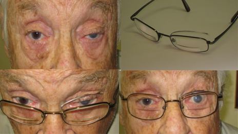
However, acquired ptosis is most commonly caused by dehiscence or disinsertion of the levator aponeurosis, causing a disconnection between the eye lid and the elevating muscles.

This usually occurs in elderly patients, but can sometimes affect younger contact lens users.

L ptosis from levator dehiscence - Note that when the eye is closed, the lid crease is fainter and further away from the lid margin in the left eye, compared to the right eye

L ptosis from levator dehiscence – Note that when the eye is closed, the lid crease is fainter and further away from the lid margin in the left eye, compared to the right eye

Patients who notice a drooping eyelid, unequal pupils, or double vision should consult with a neurologist in order to establish the correct diagnosis.

After that, treatment might include medical therapy for an underlying disorder (such as diabetes or myasthenia), surgery or even eye lid crutches:

Myasthenic patient with isolated L ptosis, demonstrating improvement with the eye lid "crutch"

Myasthenic patient with isolated L ptosis, demonstrating improvement with the eye lid “crutch”

Surgical options for trigeminal neuralgia

Trigeminal Neuralgia

Trigeminal neuralgia is characterized by recurrent short episodes of one-sided zapping, shooting, or excruciating spasms of facial pain. The pain may be so severe that the patient may cry out or visibly wince. The affected area of the face may become super-sensitive, with such minor stimulation as a light breeze, cold temperature, water from a shower, washing the face, shaving, or even eating can set off attacks of pain.

Medical Treatment of Trigeminal Neuralgia
The anticonvulsant carbamazepine is usually first line treatment.
Second line medications include baclofen, lamotrigine, oxcarbazepine, phenytoin, gabapentin, pregabalin, and antidepressants such as amitriptyline.
However these medications are not always effective and can cause side effects such as confusion and unsteady gait.

Surgical Treatment of Trigeminal Neuralgia
Surgical treatment, known as microvascular decompression, involves sub occipital craniotomy, mobilizing the offending blood vessel off the nerve, and insertion of a sponge to separate these structures.

Partial lesioning of the trigeminal nerve, either by radiofrequency ablation using a percutaneous needle or gamma knife radiosurgery is less invasive and can also be very effective:

Gamma knife for trigeminal neuralgia patient story:
20130124124455899_0001

Diabetic Neuropathy, You’d better be checking your blood sugars!

Blood_Glucose_Testing

Diabetes causes high blood sugar levels, either from lack of insulin (type I diabetes) or insulin resistance (type II diabetes).

Many diabetics develop numbness, tingling, neuropathic burning pain, or weakness that starts in the feet and progresses up the legs (“glove and stocking” pattern) from diabetic polyneuropathy.

Neuropathic pain in feet

Neuropathic pain in feet

Loss of protective sensibility from neuropathy can cause painless injuries that result in ulceration, infection and bony deformity “Charcot joints”.

Diabetic ulcer

Diabetic ulcer

Diabetic foot deformity, Charcot joint

Diabetic foot deformity, Charcot joint

There are many medications that can reduce the symptoms of neuropathic pain, such as gabapentin, pregabalin and duloxetine.

However, the only treatment that has been shown to be effective for diabetic neuropathy is improved control of blood sugar.

While many patients and their doctors rely on the glycosylated hemoglobin to measure how well their diabetes is controlled, it is important to recognize that this is an average measure of blood glucose levels, and patients with mild diabetes can have peaks (which are neurotoxic) and troughs (which can also be harmful), which average each other out, leading to a “normal” glycosylated hemoglobin level , and a false sense of security that everything is OK.

Untitled-3 copy

Blood sugars – you can have a normal “average” level, but still get nerve damage from high peaks after you eat

If you have diabetic neuropathy, we suggest that you check your finger stick glucose regularly and write the numbers down in a book or on a computer spreadsheet, so that you can review them with your doctor.

We usually suggest the following schedule to our patients:

Pick the same 2 days each week (no matter what you have eaten those days) to do the test.  On those days, check a “fasting” sugar when you first wake up (should be <130, ideally <110), a second sugar test before dinner (should be <130, ideally <110), and a third test 2 hours after dinner (should be <180, ideally <140).

Remember, although improved blood sugar control will prevent progression of your neuropathy, it won’t make it better, so start today before things get too bad!

MMC Uses New Treatment for Brain Hemorrhage

Background:

Spontaneous (non-traumatic) intracerebral hemorrhage (ICH) is a type of stroke caused by bleeding into the brain.

ICH typically causes sudden headache with stroke like symptoms.

ICH accounts for 20% of all strokes.

The mortality rate of ICH is 40%, and more than 20% of survivors are left with significant disability. Death and disability is more common with larger or expanding bleeds.

ICH is more common in patients who are taking blood thinners like warfarin (Coumadin).

Warfarin associated ICH is more likely to expand from continued bleeding leading to a worse outcome or death, so it is very important to reverse the effects of warfarin as quickly as possible in these patients.
Untitled-1 copy

An injection of Vitamin K will reverse the effects of warfarin, but it takes 12-24 hours to work.

Fresh frozen plasma (FFP) will reverse the effects of warfarin more quickly than vitamin K, and this has been the standard treatment for warfarin associated ICH, but FFP is a blood product that requires cross-matching and has to be thawed out and administered slowly.

Studies have shown that many patients treated with Vitamin K and FFP have not yet had the effects of warfarin completely reversed even by 24 hours after admission, and are still therefore at risk for hematoma expansion, death and disability.

Prothrombin Complex (PCC) is a new concentrated plasma protein product that can given by immediate intravenous injection, and along with Vitamin K and a small amount of FFP reverses the effects of warfarin within minutes, preventing ICH expansion and improving survival in ICH patients.

Untitled-2 copy

We now have PCC (Profilnine) on formulary at Monmouth Medical Center for this situation.

Case Report
A 75 year old hypertensive woman who was taking warfarin because of previous stroke was brought to our emergency room with headache and confusion. A brain CT showed ICH and her INR was modestly elevated at 1.7.

Baseline brain CT showing intraventicular blood of different ages

Baseline brain CT showing intraventicular blood of different ages

She was appropriately given 10mg of Vitamin K, 25U/Kg of Profilnine and 2 units of FFP, her INR was normalized within hours, and she did very well clinically, with some evidence of ICH resolution on a follow-up CT scan 2 days later.

Follow-up brain CT, NOT WORSE, actually showing interval improvement

Follow-up brain CT, NOT WORSE, actually showing interval improvement

This favorable outcome was the direct result of great communication and cooperation between our emergency room, ER and ICU nursing staff, pharmacy and physicians with regard to this new approach for treating warfarin associated ICH.

Click here for further information about Monmouth’s fully accredited stroke program.

A Comprehensive Sleep Medicine Evaluation

Microsoft Word - davis pamphlet version 2

Sleep disorders are common and underdiagnosed. Studies have shown that initial consultation and follow-up with a fellowship trained sleep medicine physician is more likely to resolve symptoms than direct referral for a polysomnogram by a primary care physician.

If you think you might have a sleep disorder, ask your doctor for a referral to a sleep medicine specialist!

The Importance of Correct Positioning During Anesthesia

This post is provided courtesy of K. T. Weber, Drexel University College of Medicine Class of 2013:

When a patient goes under anesthesia, a highly trained team of people take over and monitor the body. This team controls breathing, circulation and many other processes, filling in very well for the patient’s brain… however, there is one function that cannot be replicated by the anesthesia team: movement. In order to undergo surgery, a patient must have their muscles relaxed, and therefore will not move away from sore or painful spots. It is normal for people to fidget, rearranging their weight to more comfortable positions, which helps to prevent bed sores (decubitus ulcers) as well as preventing minor nerve compression.

Decubitus (Pressure) Ulcer

Decubitus (Pressure) Ulcer

Decubitus (or “pressure”) ulcers can be a devastating complication for patients who undergo very prolonged surgeries – Superman actor Christopher Reeve actually died from complications associated with an infected pressure ulcer. Fortunately, these wounds are very rare in patients who are only temporarily relaxed for surgery, and there are many precautions in place to prevent this from occurring.

severe-nerve-damage

Nerve Injury

However, minor nerve injury is still possible! One of the less commonly discussed side effects of having surgery, this minor nerve injury can be very disconcerting and affects a surprisingly large number of people

Nerve damage can occur in several forms based on the type and cause of injury:

The mildest form of nerve injury is neurapraxia, where the myelin sheath and support cells around the nerve are damaged (frequently by excessive pressure or ischemia), leading to poor signal conduction along an otherwise unharmed nerve. Neurapraxia improves on it’s own with time as the myelin sheath regenerates, and does not directly involve injury to the axon of the nerve.

untitled2

Neurotmesis, the most severe type of injury, occurs when a nerve is lacerated, over-stretched or very badly crushed, and the two ends of nerve are actually separated from each other.  These nerve injuries will not get better on their own without nerve graft repair surgery.

Untitled-1 copy

Axonotmesis is an intermediate type of nerve injury, where the nerve is damaged (often by crushing) but the support cells around the nerve are intact and aid regeneration, so (slow) spontaneous recovery is possible.

The most common symptom of compressive nerve injury is numbness or pain. The tingling, pins-and-needles feeling of hitting one’s funny bone is a classic example of nerve pain.  Many patients who have undergone surgery and are held in one position for an extended period of time may wake up with areas on their skin that feel extra sensitive, like a sunburn. It is understandable to be concerned by waking up with hyperaesthesia, or pain in response to something that wouldn’t normally be painful! Weakness and uncoordination is less common, and only occurs after more severe or prolonged nerve compression during surgery.

The good news for patients who wake up with an unusually sensitive area or some numbness after having surgery is that this minor nerve damage tends to improve rapidly. Neurapraxia resolves as the swelling and bruise around the nerve decreases and the cells that help conduction recover, sometimes as quickly as a few hours, sometimes as slowly as several months. If necessary, non-steroidal anti-inflammatories, other medications for neuropathic pain and splinting can help treat the symptoms of mild nerve damage. Even more severe forms of nerve  injury can recover with time – peripheral nerves grow back around 1mm per day! In the most extreme cases, without the potential to regenerate on their own with time, nerve graft repair can be discussed as an option.

Watch a video presentation on nerve injury and repair:

Of course, an ounce of prevention is worth a pound of cure. The skilled members of the operating room staff take extensive measures to protect their patients with adequate padding and careful positioning.

What causes numbness and tingling?

Numbness and tingling is one of the most common reasons for a visit to the neurologist, and it is usually the pattern or distribution of the numbness that is the key to figuring out the diagnosis:

1. Numbness in the hands:
This is most often caused by carpal tunnel syndrome (CTS). CTS is caused by median nerve compression at the wrist(s), and will usually present with numbness and tingling in one or both hands, mostly affecting the thumb, index and middle fingers (but sometimes the whole hand), worse at night or with certain wrist positions like driving, typing or holding a book, and alleviated by vigorously shaking the hand. In severe cases, there can be weakness or wasting of the muscles at the base of the thumb. Most patients can be treated conservatively with a neutral position wrist splint, but severe or recalcitrant cases will require surgery.
cts

Ulnar neuropathy at the elbow (funny bone) can also cause numbness and tingling in the hand, usually mostly affecting the ring and little fingers, worse at night or when leaning on the elbow. Severe cases can be associated with wasting of the muscles between the knuckles and clawing of the hand. Mild cases can be treated with an elbow pad, but severe cases will necessitate surgery to decompress or transpose (move) the nerve.
UNE

2. Numbness in one foot:
A plantar neuroma is caused by injury and scar tissue, and causes pain and numbness between the toes, worse with weight bearing and walking, and usually responds to a local injection, although occasionally needs surgical excision. Tarsal tunnel syndrome, sometimes thought of as the lower extremity equivalent of carpal tunnel syndrome, results from compression of the tibial nerve at the ankle, and causes pain and numbness in the sole of the foot, worse from standing and walking. Symptoms sometime improve with orthotics, but rarely necessitate surgical decompression. Numbness in one foot can occasionally be seen from lumbar radiculopathy,but then will be usually be associated with back pain and sciatica.
foot

3. Numbness in both feet:
So called “glove and stocking” or length-dependent numbness indicates numbness, tingling, and (in some cases) burning pain in the distal extremities usually from axonal polyneuropathy. The diagnosis can be confirmed by electrodiagnostic testing and/or skin biopsy. The most common cause is diabetes. Treatment is aimed at the underlying process, such as controlling the blood glucose in diabetics, scrupulous foot care to prevent painless injuries, and medications for symptomatic treatment of neuropathic pain.
neuropathy

4. Numbness in one thigh:
This is usually from meralgia paresthetica, which results from compression of the lateral femoral cutaneous nerve in the groin, and causes numbness and burning pain the the lateral thigh. It can be triggered by weight gain, weight loss or tight fitting clothes, and usually resolved spontaneously, although recalcitrant cases can be treated with a nerve block.
meralgia

5. Numbness in both legs:
Numbness in both legs that comes up on to the waste in a sensory level usually indicates spinal cord lesion, and can be of acute onset in transverse myelitis, spinal cord infarction or disc herniation, or more chronic onset from multiple sclerosis, spinal cord tumor, or vitamin deficiency.
myelopathy

6. Numbness that radiates down one arm or one leg
This is most likely radicular pain from a pinched nerve root in the neck or back.
radic

Piriformis syndrome from compression of the sciatic nerve in the buttock is rare cause of “sciatica”, and is frequently misdiagnosed as radiculopathy. Affected patients complain of pain in the buttock that radiates down the back of the leg, made worse by sitting or wearing a wallet in the affected back pocket, and relieved by standing and walking. Many patients improve with therapy and exercise, but some require nerve blocks or decompressive surgery.
piriformis

7. Numbness on one side:
Numbness that affects the face, arm and leg on the same side of the body usually indicates a brain lesion on the opposite side. Sudden onset one sided numbness can indicate a stroke. More gradual onset numbness on one side can indicate multiple sclerosis or a brain tumor. Affected patients will need to undergo a brain imaging study, and treatment will depend on the underlying cause.
stroke

Do you have a sleep disorder?

5-tips-for-fighting-jet-lag-4

Excessive daytime sleepiness is often a symptom of an underlying sleep disorderClick here to take an on-line test to see how sleepy your are.

sleepdisorder

Obstructive Sleep Apnoea and Snoring
  • Sleep apnea is estimated to be prevalent in about 5% of the adult population.  It is characterized by partial or complete obstruction of the upper airway with continued respiratory effort.  As an individual sleeps, one’s upper airway may collapse, leading to interruptions in breathing known as apneas.  As these continue throughout the night, one has to wake up repeatedly to breathe normally.  These “micro-arousals” from sleep lead to a non-restful sleep period and sleepiness during the day.  The typical symptoms of sleep apnea are excessive daytime sleepiness, loud snoring, and having apneas that are witnessed by a bed partner.  Obesity is a predisposing risk factor.  Sleep apnea itself is a risk factor for uncontrollable hypertension, heart attack, and stroke.  It is treated by using a positive pressure breathing apparatuses that work as a “pneumatic stent” to prevent the airway from collapsing.
insomnia

Insomnia

  • Insomnia is a highly complex sleep disorder that can manifest as excessive sleepiness.  Insomnia is very often secondary to other medical issues, other sleep disorders, or most commonly, stress and anxiety.  Obtaining a thorough medical history, including a discussion of stress and anxiety, is essential in the proper diagnosis and treatment of insomnia.
RLS

Restless Legs

  • Restless leg syndrome can prevent sleep, and therefore result in sleepiness during the day.  It is characterized by an urge or desire to move or kick legs while trying to fall asleep.  People who have this syndrome often need to get up and walk around to relieve this sensation.  It can be treated with a variety of medications.  Interestingly, iron deficiency can sometimes cause RLS and supplementation with iron in these people can often alleviate the symptoms.
narcolepsy

Narcolepsy

  • Narcolepsy is a disorder of inappropriate intrusion of sleep into being awake.  It is typically diagnosed in individuals in their 20’s.  The popular misconception of narcolepsy is somewhat misleading.  Most people with narcolepsy have an unshakeable desire to sleep and often doze off in inappropriate settings.  However, most people do not simply collapse on the street, asleep – as popular depictions of narcolepsy suggest.  Narcolepsy is often accompanied by symptoms of sleep paralysis, a sensation of being paralyzed after waking up for about a minute, hallucinations when falling asleep or waking up, and cataplexy.  Cataplexy is a unique symptom of narcolepsy where individuals lose muscle tone when they are surprised.  It is somewhat rare, but is diagnostic of narcolepsy

The treatment of sleep disorders can markedly improve a patient’s quality of life as well as reduce cerebrovascular and cardiovascular risk.

A referral to a sleep specialist is highly recommended to evaluate for these disorders.

A polysomnogram is usually necessary to diagnose sleep disorders. The sleep lab is part of the hospital, but is an outpatient procedure during which we watch you sleep for the night.  There are electrodes attached to the head, next to the eyes, on the chin, and both legs.  There are also heart leads, a pulse oximeter, and a belt around the chest and abdomen to measure work of breathing.  Despite how it sounds, most patients fall asleep normally.

This post is provided courtesy of Dr Matthew J. Davis, Fellowship Trained Sleep Neurologist.

Jet Lag

This post is provided courtesy of K. T. Weber, Drexel University College of Medicine Class of 2013:

Most travelers are familiar with jet lag. Daytime sleepiness, difficulty focusing, uncoordination, digestive issues and immune system malfunctioning can all plague those who have ventured far from home. Nearly every travel website promises advice for curing jet lag – get outside in the sunshine, try to keep yourself awake, exercise in the evening and so on. But does any of the advice work? … and why?

untitled23

Jet lag (“desynchronosis” or “circadian dysrhythmia”) is the result of moving rapidly through multiple time zones. It is caused by disruption of the body’s natural circadian rhythm, specifically the release of melatonin by the pineal gland in response to light and dark patterns. Traveling through more than five time zones shifts a person far enough off his or her normal activity cycle that waking and sleeping are affected. When sleep gets affected, concentration and cognitive performance quickly decline. Traveling west tends to have less jet lag, because it stretches the cycle out rather than shoving the circadian rhythm ahead in the light/dark pattern as traveling east does.


Melatonin is the link between the external environment and internal clock. It is released by the pineal gland, and light inhibits the release. Therefore it makes sense that melatonin, associated with dark, acts in favor of decreased activity and promoting sleep. Melatonin supplements (exogenous melatonin) is available from health food stores and a variety of non-prescription sources, and preliminary data suggests it decreases jet lag when taken shortly before bedtime. However, there is relatively little data to support the efficacy of taking melatonin supplements for sleep in other settings, partly because the source and dose of melatonin in supplements are not standardized or regulated. Ramelteon, a prescription melatonin agonist, is used for treating insomnia and may offer some benefit in the transient circadian rhythm disruption of jet lag.

Normal Sleep Regulation

Normal Sleep Regulation


Popular remedies for jet lag include:

Drinking coffee to stay awakeCaffeine has been shown to help reduce the effects of sleepiness associated with jet lag. However, it does not improve sleep, and in a slow-release formula, caffeine can actual generate less restful sleep. So a cup of coffee won’t cure jet lag, but it might help provide some alertness to counteract it. Similarly to caffeine, alcohol disrupts the sleep cycle by disrupting sleep architecture. Alcohol often makes people feel sleepy, but it impairs their rest. So both caffeine and alcohol can further upset the sleep schedule!

Getting plenty of sunshineLight exposure is the main connection humans have to the 24 hour day, so it makes sense that getting light (especially at the right time of day) would help resynchronize the biological clock. This clock can be moved about one hour closer to normal each day using light cues. For those who traveled east, and cannot fall asleep until much later in the night, it is important to get exposure to bright light in the morning. For those who traveled west and are sleepy too early in the evening, it is important to get bright light in the late afternoon, and dim light in the mornings.

and finally, refusing to change your schedule. For individuals traveling more than 5 time zones for a 2 day stay, studies have found that those who maintain their “home” sleep schedule and do not try to adjust fare better than those who try to adapt to local time! Of course, one of these studies also found that people prefer trying to adapt, for social and convenience reasons.

Whatever inspires you to next get jet-lagged, keeping in mind the physiologic basis will give you the best tools for recovering. Happy travels!