MS, Tysabri and PML

ms4

xx

The disease:

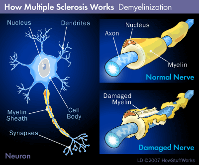
Multiple sclerosis (MS) is an auto-immune disease characterized by episodes of multi-focal inflammation and demyelination of the brain and spinal cord, leading to recurrent and unpredictable neurologic compromise (relapses or exacerbations), usually alternating with periods of disease inactivity (remissions).

ms1

xx

The drug:

Tysabri (natalizumab) is a monoclonal antibody that binds to the cell adhesion molecules involved in the white blood cell movement from the blood stream into the central nervous system across the  “blood-brain barrier”.

Keeping these cells out of the brain and spinal cord can help prevent the immune-mediated inflammation and demyelination that leads to clinical relapses in multiple sclerosis.

Studies have shown that patients taking Tysabri have a 64% reduced risk of disability progression and  >80% fewer exacerbations (relapses) compared to placebo.  More than 1/3 patient who take the drug are clinically free of disease activity.

Tysabri is administered by iv infusion once  a month.

xx

The problem:

ms2

More than 50% of people have been infected with the JC or John Cunningham) virus (JCV), most during childhood or adolescence, often with no symptoms at all or just a minor febrile illness.

Once infected, the virus then lies dormant in the central nervous system, like a Tojan horse, totally inactive and innocuous.

However, if the infected person becomes immune suppressed, for example from HIV infection (AIDS) or from taking a immune presupposing medication, the virus can become reactivated and lead to a very serious brain infection known as Progressive multifocal leukoencephalopathy (PML).  PML leads to large confluent areas of brain infection and demyelination (below), causing disability and death,

ms3

Large confluent areas of demyelination in PML.

When a few Tysabri patients developed PML during the initial clinical trials, the FDA temporarily pulled the drug, but then re-introduced it with more careful monitoring (the TOUCH porgram).

xx

I’m taking Tysabri, what’s my risk of PML?

There have now  been >350 cases of PML in MS patients taking Tysabri, and this constitutes an overall risk of about 1.5 cases per 1000 (or 0.15%) of those taking the drug.

The risk is higher for patients who have already been been exposed to (and test positive for) JCV, have taken other immunosupressive drugs, or have taken Tysabri for longer times:

ms5

So, if you take Tysabri but test -ve for JCV, your PML risk is 0.07 per 1000, or 0.007% or 1 in 14,000.

Even if you test positive for JCV but haven’t taken prior immunosupressive meds (like azathioprine, methotrexate or mycophenolate) your PML risk is only 0.6 per 1000 or 0.06% or 1 in 1,700.

xx

So, what should I do?

If you take Tysabri, you should know your JCV status.  If your negative, you should get re-tested every 6-months since there are false negative results and some people do seroconvert every year.

If (or once) you test positive, you don’t need any further blood tests, but you should carefully weigh the risk benefits of continuing to take Tysabri beyond 2 years, particularly if you have had prior exposure to other immunosupressive drugs.

ms6

Click here to find out more about Tysabri and PML from the MS society.
Correct clinical diagnosis of dizziness in the ER could save $$ billions

vertigo_dizziness-150x150
We have already blogged about the danger of escalating health costs in the USA.

The cost of emergency room visits for severe dizziness has grown exponentially in recent years, topping $3.9 billion in 2011 and projected to reach $4.4 billion by 2015.

Investigators from Johns Hopkins estimate that half a billion a year could be saved immediately if emergency room physicians stopped the routine and excessive use of head CT scans to search for stroke in dizzy patients, and instead used simple bedside physical exams to identify the small group of patients that truly needs imaging.

Click here to find out more about the Johns Hopkins study.

Click here to find out more about the hidden dangers of unnecessary CT scans.

Once they understand how inner ear disease can cause vertigo and nystagmus, any physician can use our simple clinical scheme to distinguish inner ear problems from more serious and rarer central causes like stroke in dizzy patients.  Click here to find out how.

A ripping yarn – a tale of cervical artery dissection

Case Summary:
This 46-year-old woman was healthy except for a history of occasional migraine headaches and cigarette smoking. On the day of admission she had fallen down a short flight of steps carrying a heavy box. About 2 hrs later she complained of some neck pain.  Then later that evening developed abrupt onset left sided weakness. She arrived at the emergency room within 1.5 hrs of the onset of weakness. On examination, she was alert, but she had a right gaze deviation (she wouldn’t look to the left side) and the left side was completely paralyzed. She had a normal brain CT scan.

The stroke team was notified, and she was given intravenous thrombolytic (“clot busting”) drug therapy within 1/2 hr of her arrival at the hospital and 2 hrs since the onset of her symptoms.

Carotid ultrasound subsequently showed no flow in the right internal carotid artery, and carotid arteriography subsequently showed near occlusion of the artery from an arterial dissection (see image below, red arrow):

dissection2

xx

What is cervical artery dissection?

Cervical artery dissection is caused by bleeding inside the wall one of the major arteries in the neck.

This process is thought to be triggered by local injury to the inside layer of the vessel wall.

Cervical artery dissections occur from blunt trauma:

dissection4

Cervical artery dissection can also occur after minor trauma, particularly in someone with a genetic predisposition:

dissection5

xx

What diseases predispose to arterial dissection?

There are some specific syndromes such as Marfan syndrome, Pseudoxanthoma elasticum and Ehlers- Danlos syndrome type IV that are associated with a weakness in the arterial wall making an arterial dissection more likely:

dissection6

dissection7

dissection8

In other cases, the specific cause of arterial weakness is unknown, but there is ongoing research to try to identify genetic links.

xx

What happens after a cervical artery dissection?

Symptoms can be caused from the damaged arterial wall itself (local symptoms) and some affected patients will later develop strokes.

Local symptoms include neck pain, unusual headache and/or Horner’s syndrome.

dissection9

L Horner’s syndrome (small pupil and drooping eyelid) caused by damage to the sympathetic nerve fibers in the arterial wall from carotid dissection. Click here to find out more about Horner’s syndrome and other causes of unequal pupils.

xx

What about stroke?

Stroke symptoms only occur in 25-30% dissections and can occur several days after the neck trauma and/or onset of local symptoms.

The arterial dissection narrows the space inside the blood vessel (the lumen), so less blood flow gets to the brain:

dissection1

A carotid artery dissection with blood clot inside the arterial wall (left) leading to narrowing of the vessel lumen and less blood flow (right).

Cervical arterial dissections can also cause stroke when pieces of blood clot break off and move with the blood flow only to block small arteries further inside the brain (cerebral thromboembolism), or if the dissection tracks across (and blocks off) an arterial branch (see below):

dissection3

xx

How is arterial dissection diagnosed?

Magnetic resonance imaging is probably the easiest way to make the diagnosis:

dissection10

MR angiogram (left) showing tapered occlusion of the left internal carotid (white arrow) from dissection. Fat suppressed T1 weighted MR axial image through the dissected cervical artery (right) showing bright blood within it’s wall (black arrow) from dissection.

xx

How is it treated?

In most cases the arterial dissection ultimately heals on its own without any surgical intervention.  There has been some controversy surrounding the use of anticoagulant vs anti-platelet drugs for stroke prevention after cervical artery dissection, but most current data favors the use of the anti-platelet drug aspirin:

dissection11

Of course, for patients presenting with symptoms of acute stroke, throbolytic therapy is also an option, and can improve outcome without increased risk in stroke from dissection:

dissection12

Click here to find out more about cervical artery and dissection and stroke.

Click here to find out what to do if you think your having a stroke.

Click here to find out more the certified stroke center at Monmouth Medical Center.

Lactic Acid gone to your head?

ataxia
D-Lactate Encephalopathy and Periodic Ataxia
Posted by Michael Twomey, Drexel MSIV, Class of 2014

Remember that pain in your side during a long hard workout? That’s the buildup of lactic acid or L-lactate which is a way for your body to burn energy when it there isn’t much oxygen around (like when you think it’s a good idea to run 5 miles). This feeling, although uncomfortable, won’t make you loopy–it’s twin brother, however, has no such qualms.

D-lactate is just like normal old lactic acid, only completely different. In fact, it is the mirror image of the molecule that causes us such grief. For those of us who have forgotten organic chemistry, or would rather walk on broken glass than take such a torturous class, think
of it like a pair of gloves. L-lactate would be your left hand glove and D-lactate the right. They look like the same darn thing from a distance (made out of the same leather and thread, and stuffing), but no matter how you rotate them, you could never get them to match up.

FIGURE 1

Our bodies happen to be finicky. Like the 7 year old fussy eater we all know and (hopefully still) love, the human body can only make and break down L-lactate. Thus, under factory settings, we all have an undetectable level of D-lactate in our blood. Bacteria, however, are wired a bit differently (shocking, I know). Some can take the same sugars that we eat, turn them into D-lactate, and when those buggers are in your intestinal system–this form of lactic acid can enter our blood stream.

Lactobacilli (the bad guys)

It just so happens that high levels of D-lactate effects the brain just as much as it does the rest of our bodies. We aren’t exactly sure the why or how this compound acts on our central nervous system, but it can cause anything from making you unbalanced to putting a patient in a delirious state. Case reports show symptoms of sleepiness, hallucinations, clumsiness, blurred vision, disorientation, dizziness, lethargy, excessive irritability, and abusive behavior. All of which can last up to a few days! The gait can be very unsteady (ataxic) during each episode, and this syndrome is one of the causes of “periodic ataxia”.

To make the diagnosis these patients also had an elevated amount of acid in their blood (something normal Lactic Acid can do as well) and high levels of D-lactate.
So can you blame the next time you trip over the doormat on bacteria? Probably not.
Fortunately the only reports of this syndrome have been found in people with extensive small intestine surgery. It turns out that the bacteria who can make D-lactate tend to live in our large intestine where they almost never see large amounts of sugar. Normally our small intestine is long enough to absorb all of those nutrients and leaving them to digest your Mexican meal instead. People with short bowel syndrome, however, should be aware of this possibility and eat appropriately. Otherwise having meals high in simple sugars could cause you to end up in the hospital with some strange behaviors and a nice long course of antibiotics!

Foreign Accent Syndrome – Their “Problem” or Yours?

FAS3

Foreign accent syndrome (FAS) is a rare condition which causes affected patients to suddenly speak their native language in a foreign accent.

Cases of FAS were reported as early as 1900.  However, one of the best known historical cases is “Astrid L”, a Norwegian woman who suffered a traumatic brain injury from shrapnel during a WW2 air raid in 1941.  She survived, but found herself mispronouncing vowels in such a way that she seemed to have a German accent, leading to social isolation and stigmatization for the remainder of the war.

Since then, there have been about another 60 FAS cases reported in the literature and media, mostly in patients who have suffered acute neurologic events such as strokes, multiple sclerosis and head injury.

Unlike most neurologic syndromes, FAS has not been localized to a lesion in a particular brain area.

The only thing that can be said is that most affected patients have lesions affecting the dominant hemisphere in or around known language areas.

FLS

Brain imaging studies from a FAS patient: The MRI (left) shows enlargement of the Sylvian fissure from atrophy of the left temporal lobe. The PET scan (right) shows focal hypometabolism in the left temporal lobe.

Many affected patients were initially mute, then developed FAS as they recovered from a non-fluent aphasia:

There are also some cases of FAS that have developed after minor neurologic events, or even without any clearly identifiable neurological cause at all.  Some of these patients have had normal brain imaging, suggesting that the problem can be functional or psychogenic.

This is all further complicated by the fact that different listeners can perceive different accents in a single speaker.

The video clip is a patent with FAS syndrome after brain injury from hemiplegic migraine.  She is said to have a Chinese accent.  Does it sound Chinese or just slurred to you?

The table below is from a FAS case report, where the affected patient’s “foreign accent” was obviously described very differently by observers.

FAS2

This suggests that FAS may not be a true syndrome after all, but simply a listener-bound epiphenomenon.

What does this mean?

Well, we have already explained that most FAS patients have some kind of speech or language problem that changed how they speak.  That explains the association with lesions in the dominant hemisphere.   However the “foreign accent” may actually just something perceived by the listener – the variability of perceived accents is explained by the fact that listeners have different experiences with languages other than their own.

In other words FAS may not be a true syndrome, but simply an epiphenomenon that exists only in the ears of the beholder.

Deep Brain Stimulation for Essential Tremor

Essential-tremor-is-characterized-by-sporadic-and-postural-tremor-on-hands

We have already blogged about “benign” essential tremor.

xx

Here is a recap of the patient featured in one of those previous posts. He has a long history of worsening essential tremor, which has not responded to oral medications, and has made it impossible for him to hold a cup or write with a pen:

xx

He decided to undergo deep brain stimulation, a procedure where tiny electrodes are placed into deep nuclei inside the brain. When these electrodes are activated, they cause an interruption in the brain circuits which cause tremor:

xx

Here he is, after surgery, with the electrodes switched off:

xx

Here he is with the electrodes to both sides of the brain switched on, see how much better his tremor is, particularly on the right side:

xx

He can write and hold a cup for the first time in >10 years.

These same pathways can be inactivated by gamma knife radiosurgery in tremor patients who cannot undergo deep brain stimulation because of blood thinners, dementia or some other medical problem.

Poor migraine control leads to chronic daily headache

Kuwait-invasion-oil-fire-0001

Many patients manage their infrequent migraine headaches with triptan medications, such as sumatriptan.

We call these “abortive” medications – you take them as needed whenever you have a headache to make it go away.

These medications set off an “explosion” of chemicals inside the brain, “extinguishing” the migraine just like an explosion of dynamite can put out an uncontrolled oil rig fire:

However, sometimes, that “chemical explosion” doesn’t put the fire out completely, and it comes right back.

We call this “rebound” headache, and we have already blogged about how taking too much abortive medication (including over the counter medications like Excedrin Migraine) for migraines can lead to  headache all the time, chronic daily headache, because of analgesic rebound.

Data from a new study has recently confirmed this:  The large American Migraine Prevalence and Prevention (AMPP) study showed that patients with very poor headache control were 4 times more likely to progress into chronic migraine during the following year than those with better control.

Clearly, poor headache control leads to more and more headaches, presumably because of analgesic rebound.

The solution?  Obtaining more sustained migraine control by starting a daily preventative medication for migraine like topiramate, valproic acid or botulinum toxin.

If your headaches are getting more frequent or out of control, seek the help of a board certified neurologist sooner rather than later!

What’s best for stroke prevention, aspirin, clopidogrel or both?

We have already blogged about the benefits of anti-platelet agents in stroke prevention.

We haven’t talked about how they work, or which one(s) are best.

Platelets are an important component of blood clotting (or hemostasis) – the normal process that stops us bleeding and facilitates healing after a cut or other injury:

This same platelet led coagulation cascade can lead to blood clot formation inside intact but diseased arteries laden with atheroma – the result of years of hypertension, high cholesterol and smoking:

These small blood clots then break off and travel down the artery causing (in the case of a cerebral blood vessel) a TIA or stroke.

There are three currently available  anti-platelet medications  – aspirin, modified release dipyridamole with aspirin (Aggrenox) and clopidogrel (Plavix) which inhibit platelet activation and aggregation.

asa

Comparative studies have shown that all three drugs reduce the risk of ischemic stroke in high risk patients who have had a previous stroke or TIA.  Aspirin is the cheapest of the three.  Aggrenox is more effective than aspirin alone, but can cause troubling headache in some patients.  Clopidogrel can be used in patients who are allergic or immune to aspirin.

Because these drugs work in different ways, investigators have asked whether combining them might be even more beneficial.

The initial studies said No.  The MATCH (2004), CHARISMA (2006) and SPS3 (2012) trials all showed that long term use of the combination of aspirin and clopidogrel failed to reduce the risk of major vascular events and also led to significant increased life-threatening bleeding complications (mainly intracranial and gastrointestinal) compared with either drug alone.

match

However, new data from the CHANCE study presented at this year’s international stroke meeting shows that a short course of combined treatment might be helpful:

The study enrolled 5170 patients who had suffered a TIA or minor stroke within the previous 24hrs, randomly assigned to one of two treatment groups:  The first group received aspirin (75-300 mg one-day loading dose followed by 75 mg/day).  The second group received the same aspirin regimen plus clopidogrel (loading dose of 300 mg followed by 75 mg/day) for 21 days, then just the clopidogrel alone after that.

The study showed that the 90d stroke incidence was lower in those who received both aspirin and clopidogrel.

The risk of hemorrhagic stroke and other severe bleeding was the same in the two groups.

In other words, short term combination anti-platelet therapy might be more effective in preventing stroke in this high risk TIA and minor stroke group, and this is something we will be offering patients seen in Monmouth’s innovative TIA Rapid Evaluation Center.

Robots helping dementia patients live independently

r2d2

We have already blogged about the Alzheimer’s epidemic.

There are already more than 5 million affected patients, and Alzheimer’s is now the 6th leading cause of death in the US.

 Caregivers spend an average of 70 to 100 hours per week providing care to an affected family member.

Alzheimer’s patients cope better in familiar surroundings.   They get worse more quickly when socially isolated.   It is more cost effective to keep affected patients at home for as long as possible, avoiding expensive residential care.

However, many caregivers need to go to work, and cannot be at home with their affected family member 24/7.

New programs using robots to provide social contact and even supervision for Alzheimer’s patients on their own at home may provide a cost effective solution to this problem.

Robot “pets” have already been used to encourage emotional behaviors for socially isolated dementia patients.

Scotland’s National Health Service  is putting robots into the rural homes of some dementia patients in a pilot scheme to help them to continue to live independently.

A relative or carer – potentially hundreds of miles away – can drive the machine around the house to check that everything is all right. The pair can also have a chat through a two-way video call system.

The robots are about 5ft tall, on wheels and have a TV screen instead of a head.

Giraff

A relative or carer can connect to the robot with a computer from any location. Their face will appear on the screen allowing them to chat to the other person.

The operator can also drive the robot around the house to check that medication is being taken and that food is being eaten.

Find out more about this innovative program.

Ongoing studies are showing that robots can provide affordable personalized cognitive stimulation, motivation and companionship to dementia patients, and potentially keep them living independently longer.

Find out more about caring for Alzheimer’s patients at home.