Cerebral aneurysms

sah2

Cerebral aneurysms are caused by an area of weakness in the wall an artery, which leads to a bulge or pocket, which can then rupture causing bleeding inside the brain, subarachnoid hemorrhage (SAH).

xx

When we consider patients with cerebral aneurysms, we need to cover these 4 issues and questions:

1. Subarachnoid hemorrhage.  What can happen when an aneurysm ruptures?

2. Clipping vs . coiling.  How are cerebral aneurysms treated?

3. Unruptured aneurysms.  What to do about if an aneurysm is identified on imaging study done for an other reason?

4. Screening.  What should you do if a family member has had an aneurysm or SAH?

xx

Subarachnoid hemorrhage (SAH)

sah1

SAH usually presents with a thunderclap headache, which is the sudden onset of the worse headache of your life.  There may also be vomiting, light sensitivity and a stiff neck.

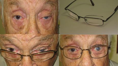
SAH patient with L IIIn palsy (left panel) and subhyloid hemorrhage (right panel).

Physical signs may include a dilated pupil and drooping eye lid (if the aneurysm arises from the posterior communicating artery PComm) and bleeding behind the eye (subhyloid hemorrhage).

sah2.2

Brain CT showing SAH (blood is bright white) tracking around the central arteries (left panel) and spinal fluid from SAH patient showing each tube uniformly red from blood (right panel).

SAH is usually seen on a brain CT scan, although in rare cases (<5%) the CT may be negative, and lumbar puncture may be needed to show blood in the spinal fluid.

Aneurysmal SAH is a very serious problem – One third of patients die before they get to the hospital.   Untreated, another third will another bleed, and two thirds of those patients will die.  Those patients who do not re-bleed can suffer delayed complications, the most serious of which is vasospasm which can lead to ischemic stroke, disability and death.

SAH patients need intensive care on a specialized neurologic critical care unit, and need to undergo further evaluation and treatment as soon as possible.

xx

How are aneurysms treated?

SAH8

Once an aneurysm is identified on cerebral angiography, there are 2 treatment options – clipping and coiling.

Clipping requires and open operation to locate the aneurysm, followed by the placement of clips around the neck of the aneurysm.

Coiling is performed through angiography – once the aneurysm has been located metal coils are inserted into the sac to cause blood clot to from and obliterate the aneurysm. 

The decision as to which treatment is undertaken is typically made by a multidisciplinary team consisting of a neurologist, neurosurgeon and neuroradiologist, and will depend on the size and location of the aneurysm, as well as the age and co-morbidities of the patient.  In general, patients who undergo coiling have quicker recovery times and the same outcome at 1 yr as patients who undergo surgery and clipping, but they have a slightly higher chance of recurrent aneurysm formation.

xx

What to do about aneurysms identified on an imaging study done for other reason?

Because of the increasing use of brain imaging, we are identifying more and more patients who have asymptomatic (or unruptured) aneurysms.  It turns out that most cerebral aneurysms never rupture.  Furthermore, there is a morbidity associated with the surgical treatment of aneurysms.   However we already know that if aneurysms do rupture and cause SAH the morbidity and mortality is very high.  So, what to do?   To answer this question, we have to study the natural history of unruptured aneurysms, and compare the probability of SAH to the risk of surgical treatment.

SAH table

Risk of aneurysm rupture (and SAH) based on its size and location.

Based on this data, you can see that the risk of rupture is very low for small aneurysms <7mm in size, and increases for larger aneurysms.

Most investigators think that the benefit for aneurysm surgery begins to exceed the risk when the aneurysm in >12mm in size.  Patients with smaller unruptured aneurysms should quit smoking, manage their blood pressure, and undergo periodic re-evaluation for aneurysm enlargement.

xx

What should you do if you have a family history of aneurysm or SAH?

Unruptured cerebral aneurysms are common and familial.  SAH is so devastating and has a poor prognosis.  So who should get elective screening for aneurysms?

For individuals with ≥2 first-degree relatives who have had  SAH, have a 8% risk of harboring an unruptured aneurysm, probably high enough to justify screening.

Individuals with only 1 affected first-degree relative have a higher relative risk of harboring an unruptured aneurysm that is only slightly higher than the general population, probably not high enough to justify screening.

Headache pills, are they hurting more than they are helping?

Headache

Headache is a common problem.  Almost 20% of men and 40% of women in the USA experience recurrent headaches.  Headache is the most common reason for a neurology office visit, the third most common cause of missed work, and the seventh most common reason for a primary health care visit.

Headaches are  broadly divided into two categories:  Primary headaches, such as migraine and tension headache, are most common.  Secondary headaches are symptoms caused by other diseases, some of them serious like a ruptured aneurysm, brain tumors, acute glaucoma or vasculitis.

Most headache sufferers are using abortive medication as needed every time they experience the symptom:

Some are taking prescription medications such as triptans or drugs containing butalbital (such as Fioricet (c) or Esgic (c)).

Many more are using over the counter medications such as acetaminophen, ibuprofen or combination pills such as aspirin/acetaminophen/caffeine (Excedrin (c)).

This strategy can work, particularly for infrequent headaches, such as once or twice a week.

cdh

However, more frequent use of these medications, particularly short-acting triptans (such as sumatriptan) and drugs containing caffeine and butalbital, can lead to rebound headache, which leads to more medication use and more headaches, culminating in chronic daily headache from transformed migraine.

cdh2

The only way to deal with this is to temporarily stop the offending abortive drug and  start a daily preventative drug.  This will lead to short period of drug withdrawal, when the headaches may get worse before they ultimately get better.  This can sometimes necessitate short term headache infusions with dihydroergotamine (DHE) or a course of steroids.

Any headache sufferer who finds themselves in this situation should consult with a neurologist or other headache specialist.

Drooping eyelids (ptosis)

Ptosis can affect one or both eyes and results from weakness affecting the muscles that raise the eyelid.

L ptosis

Left sided ptosis

Ptosis can be congenital (you are born with it), or acquired (it develops during life).

Acquired ptosis can result from a variety of problems affecting the nerves, muscles, neuromuscular junction or tendons involved in elevating the eye lids.

Neurogenic ptosis is usually unilateral, and can be caused by a lesion affecting either the oculomotor nerve or the sympathetic nerve fibers to the eye (Horner’s syndrome).

When ptosis is caused by an oculomotor nerve lesion, there is  generally also some degree of eye movement abnormality (ophthalmoparesis).  Oculomotor nerve palsy can be caused by something as simple as diabetes, but if the nerve fibers to the pupil are involved (causing a dilated pupil in addition to the ptosis and ophthalmoparesis), that is very suggestive of a compressive lesion such as an aneurysm (see below) and warrants immediate evaluation.

R ptosis, occulomotor palsy (eye is deviated down and outwards), with a dilated pupil, caused by nerve compression from aneurysm (red arrow)

R ptosis, oculomotor palsy (eye is deviated down and outwards), with a dilated unreactive pupil, caused by nerve compression from aneurysm (red arrow)

Horner’s syndrome causes mild ptosis associated with a small pupil (miosis) sometimes associated with lack of sweating (anhidrosis) on the face, and can be caused by trauma to the carotid artery, lung tumors, or strokes.

L Horner's syndrome with mild ptosis and miosis

L Horner’s syndrome with mild ptosis and miosis

Myasthenia gravis is an autoimmune disease that affects the neuromuscular junction, and frequently presents with fatiguable ptosis often associated with double vision and limb weakness.   The ptosis will usually get worse when the patients is tired at the end of the day (diurnal variation):

Fatiguable ptosis in myasthenia gravis

Fatiguable ptosis in myasthenia gravis

The ptosis of myasthenia can be temporarily improved with an acetylcholinesterase inhibitor medication, such as an injection of edrophonium (Tensilon), and this can used a diagnostic test.

Ptosis can also be seen in certain muscle diseases, including oculopharyngeal muscular dystrophy, mitochondrial myopathy and myotonic dystrophy.

Bilateral ptosis in a patient with myotonic muscular dystrophy

Bilateral ptosis in a patient with myotonic muscular dystrophy

However, acquired ptosis is most commonly caused by dehiscence or disinsertion of the levator aponeurosis, causing a disconnection between the eye lid and the elevating muscles.

This usually occurs in elderly patients, but can sometimes affect younger contact lens users.

L ptosis from levator dehiscence - Note that when the eye is closed, the lid crease is fainter and further away from the lid margin in the left eye, compared to the right eye

L ptosis from levator dehiscence – Note that when the eye is closed, the lid crease is fainter and further away from the lid margin in the left eye, compared to the right eye

Patients who notice a drooping eyelid, unequal pupils, or double vision should consult with a neurologist in order to establish the correct diagnosis.

After that, treatment might include medical therapy for an underlying disorder (such as diabetes or myasthenia), surgery or even eye lid crutches:

Myasthenic patient with isolated L ptosis, demonstrating improvement with the eye lid "crutch"

Myasthenic patient with isolated L ptosis, demonstrating improvement with the eye lid “crutch”