
Posted by Ilya Grinberg:
Last week I participated in the MDA clinic and there was one patient who really stuck out in my mind. He was a 17 year old male who had Duchenne Muscular Dystrophy. He was completely wheelchair-bound and could barely move any of his extremities. This obviously meant that he was completely reliant on people to take care of him and that he could not participate in the day to day activities that a normal person could. He was also starting to have breathing difficulty and probably would need to start therapy on a ventilator soon too.
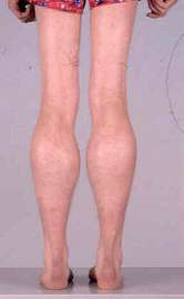
DMD is an x-linked recessive disorder that affects boys from an early age. It affects 1 out of 3600 boys so it is not too uncommon. The disease is due to a mutation of the structural protein Dystrophin which is normally found in all muscles. The protein is found on the X chromosome which explains why it is an X-linked disease. Patients are usually born normally but will start gradually developing symptoms during the first few years of life. The first observations are usually progressive muscle weakness in proximal muscles in addition to muscle atrophy. One of the key clinical signs in DMD is calf psuedohypertrophy which occurs due to fibrosis of the necrotic muscle tissue. Another common finding that presents early on is Gower’s sign, where the patient uses his arms to stand up due to weakness in the proximal leg muscles.
Click here to find out more about DMD.
Posted by John Soliman:
What is Neurology one may ask? Prior to and during medical school I feel like the exposure to the study of neurology was very limited. Interaction between neurological patients and medical professions was far and few between. I have had little encounters with the realm that lies ahead. Prior to starting the clerkship I was very intimidated due to my lack of knowledge and ignorance. I can say jokingly I barely knew how to spell Neurology. Even the basics of neurology such as anatomy was daunting usually getting hung up on learning one part or area as seen in this video. I have to say I had something in common with Pinky.
After the 4 weeks of neurology I have realized that neurology covers a broad realm of knowledge and information on the central and peripheral nervous system.
During my clerkship I was lucky enough to be exposed to many patients encountering a lot of this medical conditions and problems. I was able to identify and correlate symptoms with disease states and vice versa.
The most memorable experience was the MDA clinic. I was able to meet and was integrated into the care of a lot of the Myotonic dystrophy patients. I was able to see hear their day to day life experience, talk to their care givers and be able to help with their care. After seeing patients like this it really brought “problems” Into perspective. The amazing thing was the broad range of how these medical conditions affect each individual differently.
I had the opportunity to see two brothers who are both affected with Myotonic Dystrophy. One brother can walk, talk and act normal with minimal weakness while the other was wheel chair bound. Talking to them brought home how a muscular dystrophy can affect the lives of affected individuals. Myotonic dystrophy is an autosomal dominant genetic process which means it can affect 50% of the carriers offspring. This may affect a family’s decision on having kids both mentally and psychologically. In this specific case, one of the brothers and his wife decided to adopt children due to the high risk of having a child with myotonic dystrophy. Dealing with something like this is a full time job on its own so it can be draining mentally, physically and financially on families.
Overall the experience has been great as I have gotten to see patients with medical conditions that I may not be able to see again. This clerkship has been knowledgeable and I has encountered a broad real of neurology that I never had experienced in the past.
Click here to find out more about Myotonic Dystrophy.
Click here to find out more about MDA Clinic at Monmouth.


