Plasma Exchange For Myasthenia Gravis

Posted by Christopher Orr,  Drexel University College of Medicine 2014

Ms. AB presented last week in the Neurology office with shortness of breath and weakness, and she knew it was from her myasthenia gravis.

She was already on an anticholinesterase inhibitor, but it was very apparent that she was suffering from a severe exacerbation of myasthenia gravis.

We sent her to the Emergency Room in order to be admitted so she could receive plasmapheresis in order to minimize the antibodies that were blocking the acetylcholine receptors at her neuromuscular junctions.

To give a brief history of Ms. AB’s myasthenia gravis, she was diagnosed in the Fall of 2013 when she presented with muscle weakness and difficulty breathing.  She was treated with plasmapheresis during that initial episode, and improved.

In the interim, she had also been given steroids to reduce the immune response of her autoantibodies towards her acetylcholine receptors, but this actually caused increased leg weakness, more likely from steroid myopathy than myasthenia gravis.

Unfortunately she experienced another exacerbation in December, and she was treated with intravenous immunoglobulin (IVIG).  What is interesting is that when she was treated with IVIG, her symptoms did not improve as they had done plasmapheresis.

There is limited research on the efficacy of IVIG in comparison to plasmapheresis in the literature.  A comparison study of IVIG vs. plasmapheresis waspublished by Mandaway et. al. in the Annals of Neurology in 2010 and included 1,606 patients – both therapies showed similar clinical outcomes in terms of both mortality and complications.  From a purely financial perspective, IVIG was more cost effective because of lowered length of stay and total inpatient charges.

However, a smaller study published by Stricker et. al. in JAMA in 1993 reported 4 patients who did not respond to initial IVIG treatment but later responded to plasmapheresis.  There were no definite prognostic factors mentioned that might explain why plasma exchange may be better than IVIG in certain patients.  The article stated further research was needed.

Ms. AB did present with myasthenia gravis at a later age of onset than is typically observed.  For future studies that compare IVIG to plasmapheresis, I would be highly interested in a subgroup analysis on a patient’s age and the efficacy of the 2 treatment modalities of IVIG and plasmapheresis.

When we saw Ms. AB in the hospital, she was already doing much better with plasmapheresis.  In addition, we were For the future, Ms. AB would likely be discharged to a rehabilitation facility and there are considerations to start her on CellCept (mycophenolate mofetil).  It would be preferential to start the patient on CellCept as an immunomodulatory drug to decrease the autoantibodies against her acetylcholine receptors and reduce her need for plasmapheresis.

I chose to write a reflection on Ms. AB for 2 reasons.  First, she and her husband are both very kind people, and it is a pleasure to see her improve.  Second, I love technology in medicine and healthcare.  When we saw Ms. AB’s plasmapheresis treatment, it was fascinating to see the apparatus that was using centrifugal force to spin her blood and separate her plasma from the WBCs, RBCs, and platelets.  The mechanism behind performing the plasmapheresis was to take off her plasma, which had the autoantibodies, and replace new plasma with albumin.  Please look below to see a picture of a plasmaphresis apparatus and an explanation of how it works.  After my experience seeing Ms. AB, it was a pleasure to treat her and learn from her condition.

OLYMPUS DIGITAL CAMERA

Pictured above is an apparatus that is used for the plasmapheresis treatments.  Although this machine may seem very intimidating, it operates on the basis of how spinning blood can separate the blood into different components, such as the plasma, RBCs, WBCs, and platelets. To explain in a simple manner, a central venous line is obtained from the patient so blood can be brought to the machine and spun.  After the plasma is removed by the centrifugal force, the remaining components (RBCs, WBCs, and platelets) is added with albumin and saline (a protein found in plasma) then reintroduced into the patient.

In many instances, diseases can be complicated to comprehend.  I wanted to give a better understanding of myasthenia gravis.  I hope this picture and caption that I included make the disease easier to digest.

nmj achr

The above image shows the synapse of a neuromuscular junction.  In a healthy patient, the acetylcholine can bind the acetylcholine receptor and produce a response in the muscle.  In a patient with myasthenia gravis, the antibody (shown in green) is blocking the acetylcholine receptor and preventing the acetylcholine in the synapse from reaching the muscle.  This picture is a good educational tool because it also shows how the treatment of pyridostigmine (Mestinon) can improve a patient’s symptoms.  The acetylcholinesterase (AChE, the red pac-man figure) is what degrades the acetylcholine in the synapase.  Pyridostigmine is an anticholinesterase inhibitor and impedes the red pac-man figure in the picture from working.  Therefore, pyridostigmine increases the amount of acetylcholine in the synapse that can reach the receptor and will improve the symptoms in an episode of myasthenia gravis.

Diaphragmatic Pacing and ALS

DPS-illustration 393 249

Amyotrophic lateral sclerosis (ALS), sometimes referred to as “Lou Gehrig’s Disease“, is a progressive neurodegenerative disease that affects motor nerve cells in the brain and the spinal cord ultimately leading to muscle paralysis.

The diaphragm is a large muscle that moves air across the lungs to facilitate gas exchange and oxygenation of the tissues:

In ALS degeneration of the motor nerve cells that innervate the innervate the diaphragm via the phrenic nerves ultimately leads to respiratory failure.

diaphragm

The diaphragm (purple arrows) a large muscle innervated by the phrenic nerves (green arrows).

The diaphragmatic pacing (DP) system bypasses the degenerated phrenic nerves in ALS, and provides direct electrical stimulation to the diaphragm, facilitating enhanced ventilation.

So we know we can electrically stimulate the weak or paralyzed diaphragm and make it contract.

xx

However, the real question is does this make a difference in terms of life and survival?

We have already blogged about the symptoms of nocturnal hypoventilation and benefits of NIPPV.

Here is a link to a recent summary of studies that have looked at DP for hypoventilation in ALS:

Data presented to the FDA in 2011 included 106 ALS patients who had undergone the surgery.

DP patients lived 9 months longer than a historical cohort of ALS patients with respiratory difficulties treated with NIPPV.

Unlike NIPPV, DP patients do not have to use a mouthpiece or mask.

However, the serious surgical complication rate from the procedure was 3.5%, 26% patients report mild-moderate discomfort from the electrical stimulation, and there were many technical problems including broken electrodes.

In ALS, unlike in spinal cord injury, the denervated diaphragm muscle will ultimately become inexcitable, rendering the DP system ineffective.

DP surgery costs about $20,000, compared to <$1000 for a NIPPV system.

xx

So, the jury is still out.

But before you sign up for DP,

Consider these facts:

DP is FDA approved as a medical device not as a therapy.

Many neuromuscular physicians are calling for real outcome data before widespread adoption of this invasive and expensive interventional procedure.   In response to these concerns, the Muscular Dystrophy Association (MDA) and ALS Association (ALSA) have co-sponsored a prospective study to determine whether the DP system in effective or not.

Finally, we know that ALS patient care varies at different centers around the country.  However, we don’t know how many ALS patients are getting their FVC measured or being assessed for hypoventilation appropriately.  We hope that the new MDA sponsored clinical registry will answer this question.

xx

Find out more:

Click here to find out more about hypoventilation in neuromuscular diseases.

Click here to link to a podcast about DP in ALS.

Click here to find out more about the DP system from the device manufacturer.

Click here to find out more about the DP clinical trial.

Neuromuscular respiratory failure

lungs_1
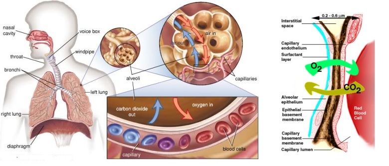
Each lung is composed of >300 million tiny membrane bound sacs of air sacs (alveoli) which if spread out would cover a piece of ground roughly the size of a tennis court.  The purpose of this giant membrane is to exchange oxygen from the air for carbon dioxide from the blood stream.

lungs1

xx

If the lungs become congested (or filled with fluid) from infection (pneumonia) or heart failure, it becomes harder to extract oxygen from the air:

lungs2

Treatment includes adding extra oxygen to the air to make this process more efficient.

cc

However, gas exchange across the alveoli can only occur if fresh air is brought into the lungs, and stale air is moved out, a process known as ventilation.  The diaphragm and muscles of the chest wall act like a giant bellows to make this happen:

lungs3

These muscles can become weak in nerve or muscle diseases such as Guillain-Barré syndrome, polio, amyotrophic lateral sclerosis (ALS), Duchenne Muscular Dystrophy and myasthenia gravis.

cc

These patients are evaluated by pulmonary function testing, which will usually show a low forced vital capacity, low cough flow, and in advanced cases, elevated end-tidal carbon dioxide level.

cc

Patients with this type of ventilatory failure do not need extra oxygen, their lungs can extract oxygen from air normally, they need mechanical assistance moving air across their lungs:

cc

lungs2

cc

Early neuromuscular respiratory muscle weakness causes nocturnal hypoventilation.  This is because the weakened diaphragm is even more inefficient when laying supine in bed with the stomach contents pressing up on it.

Nocturnal hypoventilation presents with daytime sleepiness, early morning headaches, fatigue, and impaired cognition.

5-tips-for-fighting-jet-lag-4

Click here to take an on-line test, and find out how sleepy you are during the day.  If you score 10 or higher, you might have a problem!

Nocturnal hypoventilation is best treated using a non-invasive respirator at night, either with a face or nose mask:

nippv

cc

Other patients use a negative pressure respirator vest, or cuirass, which requires the patient to wear an upper body shell  attached to a pump which actively controls the respiratory cycle:

cuirass

xx

Neuromuscular respiratory failure also leads to an ineffective cough, which in turn predisposes patients to aspiration, retention of secretions, or pneumonia.  Affected patients need to learn to use the cough assist machine when they get a minor respiratory tract infection to help them clear their secretions and prevent pneumonia:

cough assist

xx

More severe neuromuscular ventilatory failure leads to rapid shallow breathing, accessory respiratory muscle use, thoracoabdominal paradox (inward motion of the abdomen during inspiration), and ultimately high blood levels of carbon dioxide.

image2_w

Thoracoabdominal paradox – Normal (upper) abdomen moves outward with inspiration (diaphragm contraction). NM weakness (lower) abdomen moves in when patient inspires using accessory muscles.

In these cases, respiratory support is needed day and night.

Some patients can continue to use non invasive respiratory support, sleeping with a face or nose mask, and using a mouth piece intermittently during the day:

mouth

Others cannot tolerate noninvasive ventilation or have anatomic abnormalities that preclude fitting of noninvasive ventilators.  Some disease, such as advanced ALS and Duchenne muscular dystrophy, affect the upper airway muscles as well as the diaphragm, impairing swallowing and compromising airway protection from aspiration.  These patients can chose to be managed with invasive respiratory support using a tracheotomy and conventional ventilator.

trach

xx

Click here and and here to find out more about the management of neuromuscular respiratory failure.